An Anatomical Specimen of a Disarticulated Human Skull
An Anatomical Specimen of a Disarticulated Human Skull
Contained in a tiered Victorian paper box with a label to the lid reading: ‘White and Wright, Surgical Instrument Makers, 93 Renshaw St, Liverpool’
Circa 1870 – 1880
Size: 21cm high, 18cm dia – 8¼ ins high, 7 ins dia (box)
Contained in a tiered Victorian paper box with a label to the lid reading: ‘White and Wright, Surgical Instrument Makers, 93 Renshaw St, Liverpool’
Circa 1870 – 1880
Size: 21cm high, 18cm dia – 8¼ ins high, 7 ins dia (box)
Anatomy, the study of body structure, is now central to the study of disease, but it was not part of medical courses at medieval universities until the 13th century. In London the practice of dissection and the study of anatomy were encouraged as early as 1540 by an act which gave the Masters of the Barber-Surgeons the right to take each year for dissection, free, the bodies of four felons immediately after their execution.
During the 18th century disease began to be linked to changes that could be detected by examining bodies either before or after death and as a result knowledge of anatomy was increasingly in demand. From 1750 to 1850 medical students in Europe were expected to learn how to dissect well, and to learn preservation techniques and it was from real body parts that students were taught anatomy.
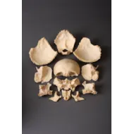
Disarticulated skulls consist of all the constituent bony parts of the human skull broken down and compartmentalised which can then be reformed like a jigsaw puzzle into a complete skull enabling the student to learn where each section and part is placed and its function.
During the 18th century disease began to be linked to changes that could be detected by examining bodies either before or after death and as a result knowledge of anatomy was increasingly in demand. From 1750 to 1850 medical students in Europe were expected to learn how to dissect well, and to learn preservation techniques and it was from real body parts that students were taught anatomy.
Disarticulated skulls consist of all the constituent bony parts of the human skull broken down and compartmentalised which can then be reformed like a jigsaw puzzle into a complete skull enabling the student to learn where each section and part is placed and its function.
An Anatomical Specimen of a Disarticulated Human Skull
SOLD
YOU MAY ALSO LIKE
